
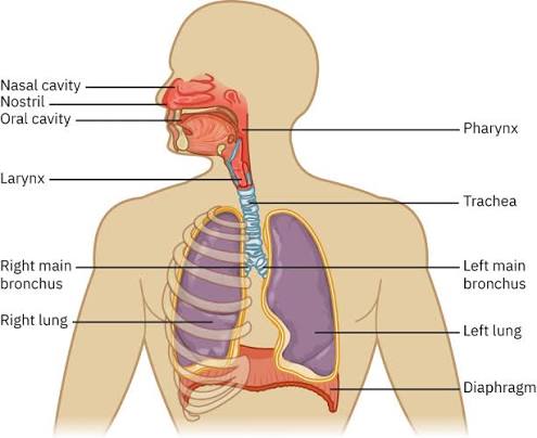
The torso is the central region of the human body, connecting the neck with the pelvis and supporting the upper and lower limbs. It contains many of the organs needed for breathing, circulation, digestion, waste removal, movement, and protection. Understanding human torso anatomy means studying not only individual organs but also the bones, muscles, blood vessels, nerves, membranes, and body cavities that support them.
In anatomical descriptions, trunk is commonly used for the central body region, while terms such as thorax, abdomen, pelvis, and back describe more specific areas. OpenStax describes the major ventral cavities of the trunk as the thoracic and abdominopelvic cavities, separated by the diaphragm.
Quick Overview of Major Torso Areas
| Area | Major Structures |
| Thorax | Heart, lungs, ribs, sternum and thoracic spine |
| Abdomen | Liver, stomach, pancreas, spleen, intestines and kidneys |
| Pelvis | Bladder, rectum, reproductive structures and pelvic floor |
| Posterior trunk | Vertebral column, back muscles and spinal nerves |
These areas are closely connected and function together rather than as isolated compartments.
The Thorax and Chest Cavity
The thorax, or chest, lies between the neck and abdomen. Its skeletal framework includes 12 thoracic vertebrae, 12 pairs of ribs, and the sternum. Together they form the thoracic cage, which protects the heart, lungs, and major blood vessels while still allowing movement during breathing.
The lungs occupy much of the thoracic cavity. Each lung is associated with a pleural cavity and pleural membranes. Between the lungs lies the mediastinum, which contains the heart, major vessels, trachea, esophagus, and other structures. OpenStax describes the lungs as occupying the pleural cavities and the heart as lying within the mediastinum.
The Diaphragm
The diaphragm is a dome-shaped skeletal muscle separating the thoracic cavity from the abdominal cavity. During inhalation, it contracts and moves downward, increasing thoracic volume and helping air enter the lungs. During relaxed exhalation, it returns toward its resting position.
MedlinePlus describes the diaphragm as the muscle that separates the chest and abdominal cavities and plays a major role in breathing. OpenStax also explains that coordinated activity between the diaphragm and abdominal muscles helps generate pressure used during functions such as urination, defecation, and childbirth.
Abdominal Anatomy and Major Organs
The abdomen lies mainly between the diaphragm and pelvis. Important structures include the liver, stomach, gallbladder, pancreas, spleen, small intestine, large intestine, kidneys, and major blood vessels.
The liver contributes to metabolism, nutrient processing, and bile production. The stomach helps digest food, while the pancreas produces digestive enzymes and hormones involved in blood-glucose regulation. Most nutrient absorption occurs in the small intestine, while the large intestine absorbs water and helps form stool. Understanding these relationships is an important part of human torso anatomy, because structures from different organ systems can lie close together within the abdominal cavity.
Abdominal Quadrants and Nine Regions
Healthcare professionals often divide the abdomen into four quadrants:
- Right upper quadrant (RUQ)
- Left upper quadrant (LUQ)
- Right lower quadrant (RLQ)
- Left lower quadrant (LLQ)
These divisions help describe organ location, tenderness, masses, and other clinical findings.
A more detailed system divides the abdomen into nine regions. The central regions are the epigastric, umbilical, and hypogastric regions. On either side are paired right and left hypochondriac, lumbar, and iliac regions. OpenStax describes both the four-quadrant and nine-region systems as standard methods for communicating abdominal location.
Peritoneal Relationships and Urinary Organs
Many abdominal structures are related to a serous membrane called the peritoneum. The parietal peritoneum lines the abdominal wall, while the visceral peritoneum covers much of the surface of many organs. Intraperitoneal organs are largely covered by visceral peritoneum and are commonly supported by mesenteries or other peritoneal folds.
Retroperitoneal structures lie posterior to the parietal peritoneum. Examples include the kidneys, adrenal glands, ureters, most of the pancreas except the tail, abdominal aorta, and inferior vena cava. NCBI’s StatPearls describes these structures as important components of the retroperitoneal space.
The kidneys are paired retroperitoneal organs that filter blood, remove metabolic waste, help regulate fluid and electrolyte balance, and produce urine. Urine moves through the ureters to the urinary bladder in the pelvis and later exits through the urethra. The adrenal glands sit superior to the kidneys and produce hormones involved in metabolism, stress responses, and blood-pressure regulation.
Pelvic Region and Pelvic Floor
The pelvis forms the lower portion of the trunk and provides structural support while protecting internal organs. The pelvic cavity contains the urinary bladder, rectum, portions of the large intestine, and internal reproductive structures.
At its inferior boundary is the pelvic floor. The pelvic diaphragm includes the levator ani and ischiococcygeus (coccygeus) muscles. These muscles help support pelvic organs and resist increases in abdominal pressure. OpenStax describes the pelvic floor as supporting pelvic viscera and contributing to urinary, bowel, and reproductive functions. Because the abdominal and pelvic cavities are continuous, they are often discussed together as the abdominopelvic cavity.
Musculoskeletal Anatomy of the Torso
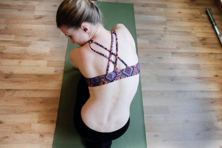
The vertebral column forms the main supporting axis of the trunk. The torso includes 12 thoracic vertebrae and 5 lumbar vertebrae, followed by the sacrum and coccyx. The thoracic vertebrae articulate with the ribs, while the larger lumbar vertebrae support substantial body weight. Intervertebral discs between most adjacent vertebrae help absorb forces and permit movement.
The 12 pairs of ribs protect thoracic organs while retaining enough flexibility for normal breathing. OpenStax confirms these vertebral and rib counts in its descriptions of the vertebral column and thoracic cage.
Important torso muscles include:
- Rectus abdominis
- External oblique
- Internal oblique
- Transversus abdominis
- Intercostal muscles
- Erector spinae
- Latissimus dorsi
- Multifidus
The abdominal and back muscles help control trunk movement, stabilize the spine, maintain posture, and support breathing. Superficial muscles often contribute to larger movements, while deeper muscles such as the multifidus are important for spinal stability.
Blood Vessels, Nerves and Body Cavities
The torso contains some of the body’s largest blood vessels. The aorta carries oxygenated blood away from the heart, while the superior and inferior venae cavae return deoxygenated blood. Pulmonary arteries carry blood from the heart to the lungs, and pulmonary veins return oxygenated blood to the heart.
The spinal cord travels through the vertebral canal, and spinal nerves branch outward to serve the trunk and limbs. The autonomic nervous system also helps regulate involuntary functions such as heart rate and digestion.
Body cavities organize and protect internal structures. The thoracic cavity contains the pleural cavities and mediastinum, while the abdominopelvic cavity lies below the diaphragm. Serous membranes, including the pleura, pericardium, and peritoneum, help reduce friction around moving organs.
Why Torso Anatomy Matters
Knowledge of human torso anatomy is useful in medicine, nursing, physiotherapy, sports science, fitness education, and biology.
Anatomical terminology helps professionals describe body regions, injuries, imaging findings, and surgical locations more precisely. It also helps students understand how multiple organ systems fit together within the same physical space.
Final Summary
The torso contains many structures essential to normal body function. The thorax houses the heart and lungs, the abdomen contains major digestive and urinary organs, and the pelvis supports lower abdominal and pelvic structures. Bones, muscles, nerves, vessels, membranes, the diaphragm, and the pelvic floor work together to support posture, movement, circulation, respiration, digestion, and organ protection.